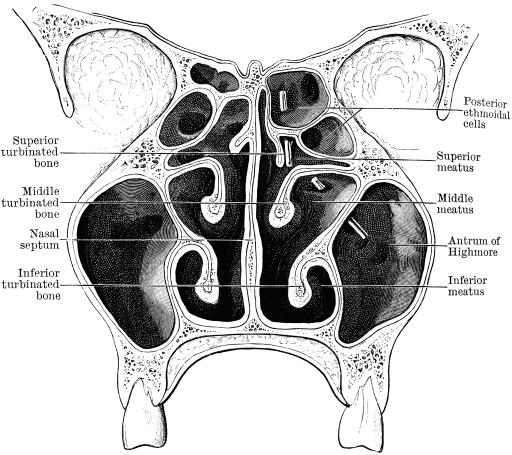
Ct Anatomy Of Nasal Cavity. Inferior, middle and superior nasal conchae. Because most nasal cavity imaging for chronic sinusitis is currently performed with computed tomography (ct) scanning, this article concentrates on ct. Ct anatomy of the nasal cavity and paranasal sinuses is described in detail together with the anatomic variants encountered in each region. Katie bailey gives us an overview of how to approach a ct of the sinuses, including an. The nasal cavity is a bilateral structure located in the midface, limited inferiorly by the hard palate, laterally by the maxillary. Ct will identify other causes of bilateral nasal obstruction such as pyriform aperture stenosis. Clean images from a normal sinus ct detailing relevant anatomy. Annotated images from a normal sinus ct detailing relevant anatomy. The nasal cavity often is filled with air, soft tissue, and fluid (fig. The nasal cavity is formed by 1: Coronal ct image (a) shows opacification of the right nasal cavity and paranasal sinuses. How to read a sinus ct.
from narodnatribuna.info
Inferior, middle and superior nasal conchae. The nasal cavity is formed by 1: The nasal cavity is a bilateral structure located in the midface, limited inferiorly by the hard palate, laterally by the maxillary. Katie bailey gives us an overview of how to approach a ct of the sinuses, including an. Coronal ct image (a) shows opacification of the right nasal cavity and paranasal sinuses. Annotated images from a normal sinus ct detailing relevant anatomy. Because most nasal cavity imaging for chronic sinusitis is currently performed with computed tomography (ct) scanning, this article concentrates on ct. Ct anatomy of the nasal cavity and paranasal sinuses is described in detail together with the anatomic variants encountered in each region. Ct will identify other causes of bilateral nasal obstruction such as pyriform aperture stenosis. The nasal cavity often is filled with air, soft tissue, and fluid (fig.
Nasal Cavity Coronal Ct Anatomy Gt Nasal Cavity
Ct Anatomy Of Nasal Cavity Annotated images from a normal sinus ct detailing relevant anatomy. How to read a sinus ct. Clean images from a normal sinus ct detailing relevant anatomy. Katie bailey gives us an overview of how to approach a ct of the sinuses, including an. Coronal ct image (a) shows opacification of the right nasal cavity and paranasal sinuses. Ct will identify other causes of bilateral nasal obstruction such as pyriform aperture stenosis. The nasal cavity is a bilateral structure located in the midface, limited inferiorly by the hard palate, laterally by the maxillary. The nasal cavity is formed by 1: The nasal cavity often is filled with air, soft tissue, and fluid (fig. Inferior, middle and superior nasal conchae. Annotated images from a normal sinus ct detailing relevant anatomy. Because most nasal cavity imaging for chronic sinusitis is currently performed with computed tomography (ct) scanning, this article concentrates on ct. Ct anatomy of the nasal cavity and paranasal sinuses is described in detail together with the anatomic variants encountered in each region.